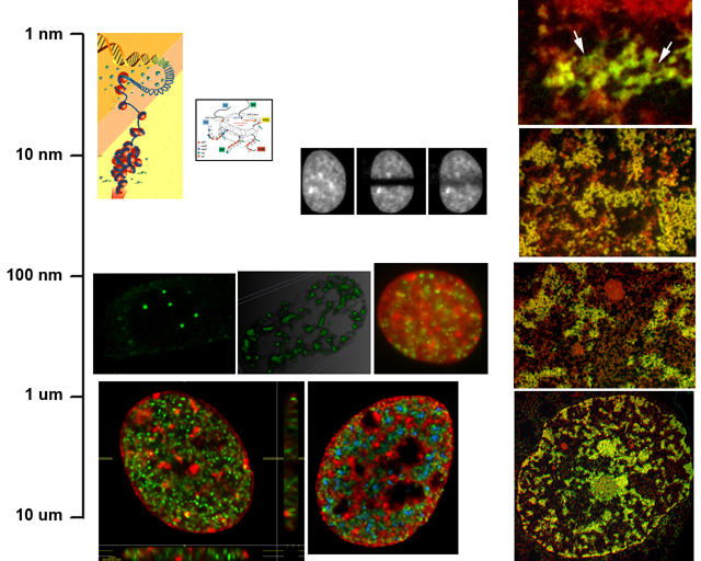
DNA within the interphase nucleus is organized into chromatin.
Chromatin is formed of a repeating subunit of 146 base pairs of DNA
wrapped around the outer surface of an octamer of four histone proteins
H2A, H2B, H3, H4. This 10 nm diameter particle is then folded into more
complex fiber structures beginning with the 30 nm fiber up to its most
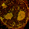
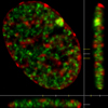
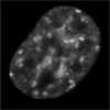
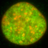
highly folded state, an approximately 700 nm diameter chromosome. The images indicate different scales of organization within the interphase nucleus. The panels on the right are electron microscopy images illustrating the different scales of organization. The images were collected by electron spectroscopic imaging (ESI). The top left panels are cartoons showing the organization of DNA into the nucleosome and nucleosome fiber. The top center image illustrates a fluorescence recovery after photobleaching image set reflecting the movement of molecules (histone H1 in this case). The center fluorescence microscopy images illustrate nuclear bodies (Gems-left, splicing factor compartments-middle, and DNA double-strand break repair foci-right). These are in the 100-1000 nm size range. The bottom fluroescence panels show whole nuclei. Nuclei are several microns in diameter |
| Copyright 2007 Michael J Hendzel, Ph.D. Department of Oncology, University of Alberta |