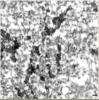
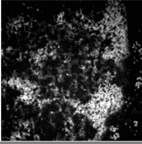
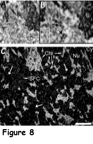
Brightfield image of chromatin and surrounding nucleoplasm. Original magnification is 20,000X
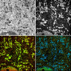
EFTEM images of chromatin and surrounding nucleoplasm. Top left-brightfield, top right-phosphorus image (chromatin and RNA), bottom left-DNA and RNA (yellow) vs. protein (red). Original magnification is 12,000X
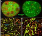
Figure4final copy.jpg
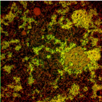
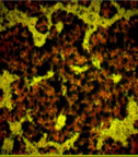
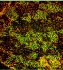
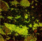
EFTEM map of chromatin and surrounding nucleoplasm. DNA and RNA is yellow, protein is red. The RNA is predominantly found in the smaller granular structures.

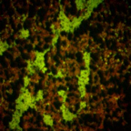
EFTEM map of chromatin and surrounding nucleoplasm. HeLa cell imaged at 20,000X.
EFTEM map at 50,000X. The small granular RNA is more evident. The chromatin concentrates in larger fibers comprised of 10 and 30 nm chromatin fibers.
eftemhelafigure.psd
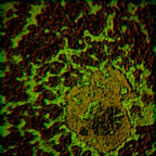
EFTEM map of chromatin at 12,000X. The orange structure in the bottom right is a nucleolus.
Net phosphorus (DNA and RNA) map of chromatin and surrounding nucleoplasm.
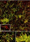
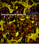
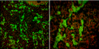
Brightfield images (top) and net phosphorus map (bottom). Chr=chromatin, IGC=interchromatin granule cluster.
EFTEM map of chromatin and surrounding nucleoplasm at 30K original magnification.
Previous image with labels.
EFTEM map of chromatin and surrounding nucleoplasm at 12,000 original magnification.
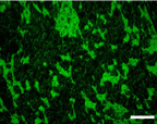
Net phosphorus map of chromatin.
Low and high magnification image of chromatin.
12,000X image of chromatin.