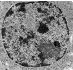
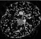
A PC12 nucleus-brightfield image.
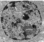
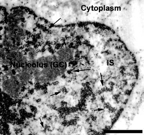
Above nucleus with labels.
A PC12 nucleus-brightfield image.
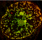
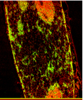
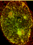
A mouse fibroblast nucleus at 7K original magnification. EFTEM showing chromatin/RNA in yellow and protein in red. The bright red spheres are nuclear bodies.
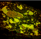
A phosphorus map of a mouse fibroblast nucleus.
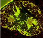
A mouse fibroblast nucleus showing a high degree of chromatin condensation. EFTEM map.
A mouse fibroblast nucleus imaged at 7,000X original magnification.
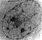
An Indian muntjac fibroblast nucleus.
A mouse fibroblast nucleus imaged at 7,000X original magnification. EFTEM map.
b7k.psd