Nuclear structure research impact

Laboratories that changed how the nucleus is understood

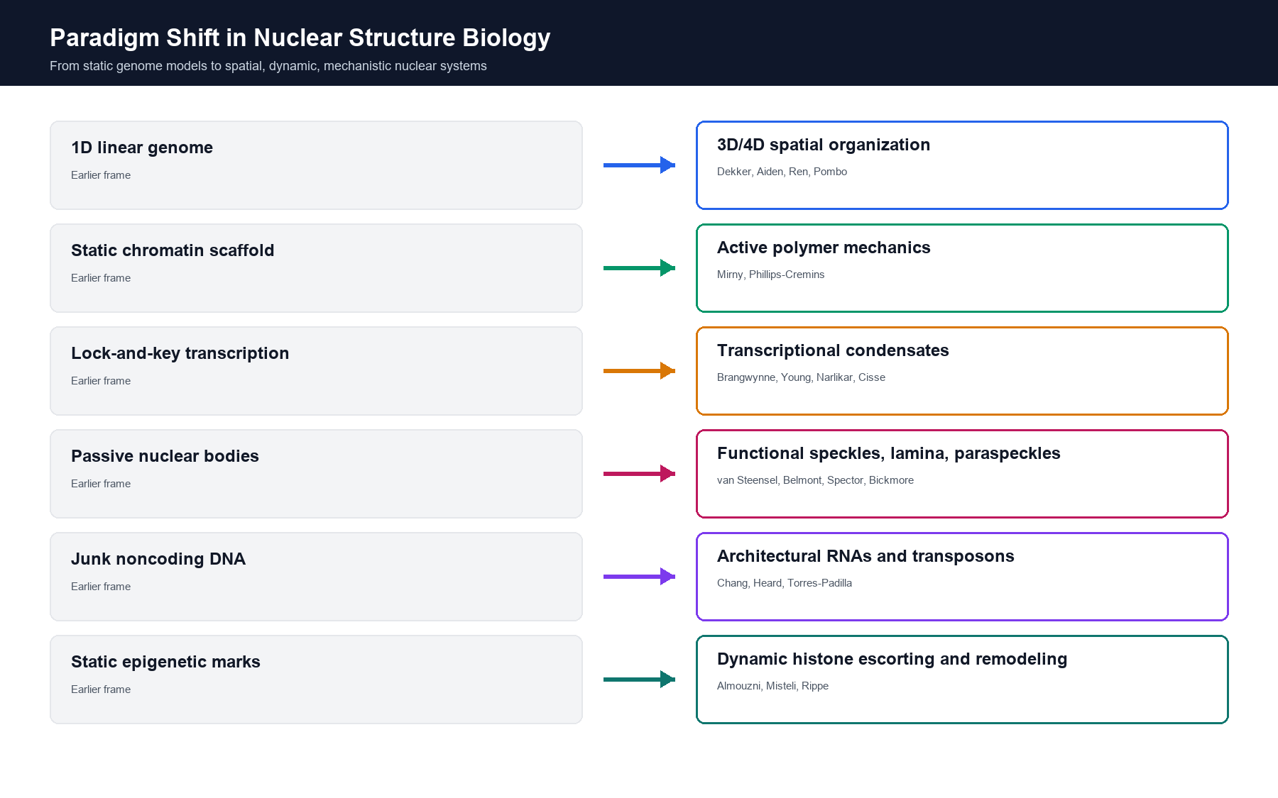
This page distills the attached research-impact analysis into a visual guide to 20+ laboratories whose tools, models, and discoveries moved nuclear biology from static chromosome descriptions toward spatial, dynamic, mechanistic systems.

Infographic showing paradigm shifts in nuclear structure biology
21

laboratory profiles represented from the document

6

research engines connecting maps, physics, imaging, and disease

3

raster infographics created for this page

1

field story: structure, mechanism, and cellular consequence

Field map

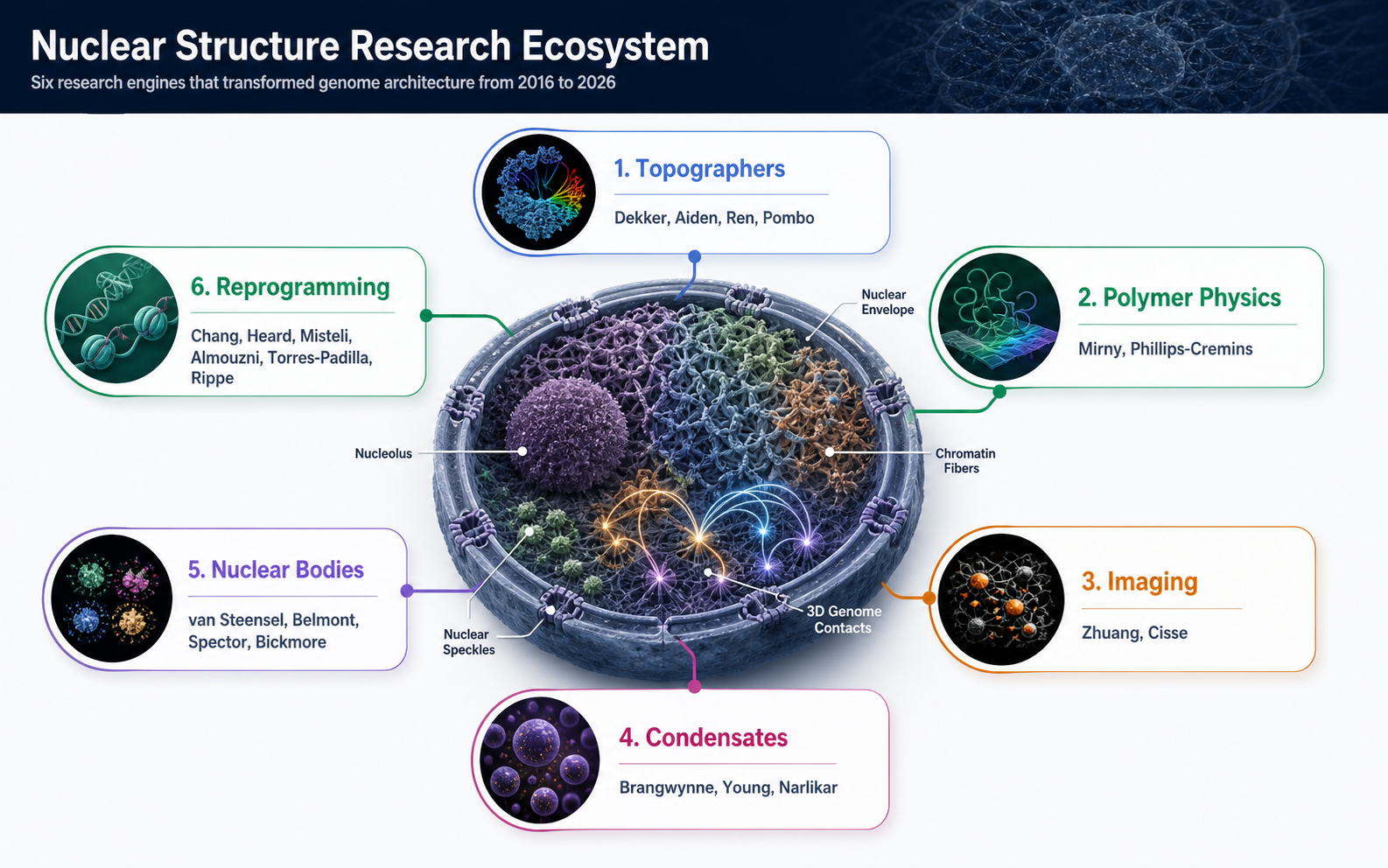
Six research engines behind modern nuclear structure biology

The laboratories cluster into complementary roles: groups that map chromosome contacts, model folding mechanics, directly image single-cell states, define condensate material rules, assign function to nuclear bodies, and connect nuclear architecture to development, aging, and cancer.

ChatGPT-generated infographic mapping leading nuclear structure laboratories into six research engines

How the field advanced

From tools to mechanisms to leverage points

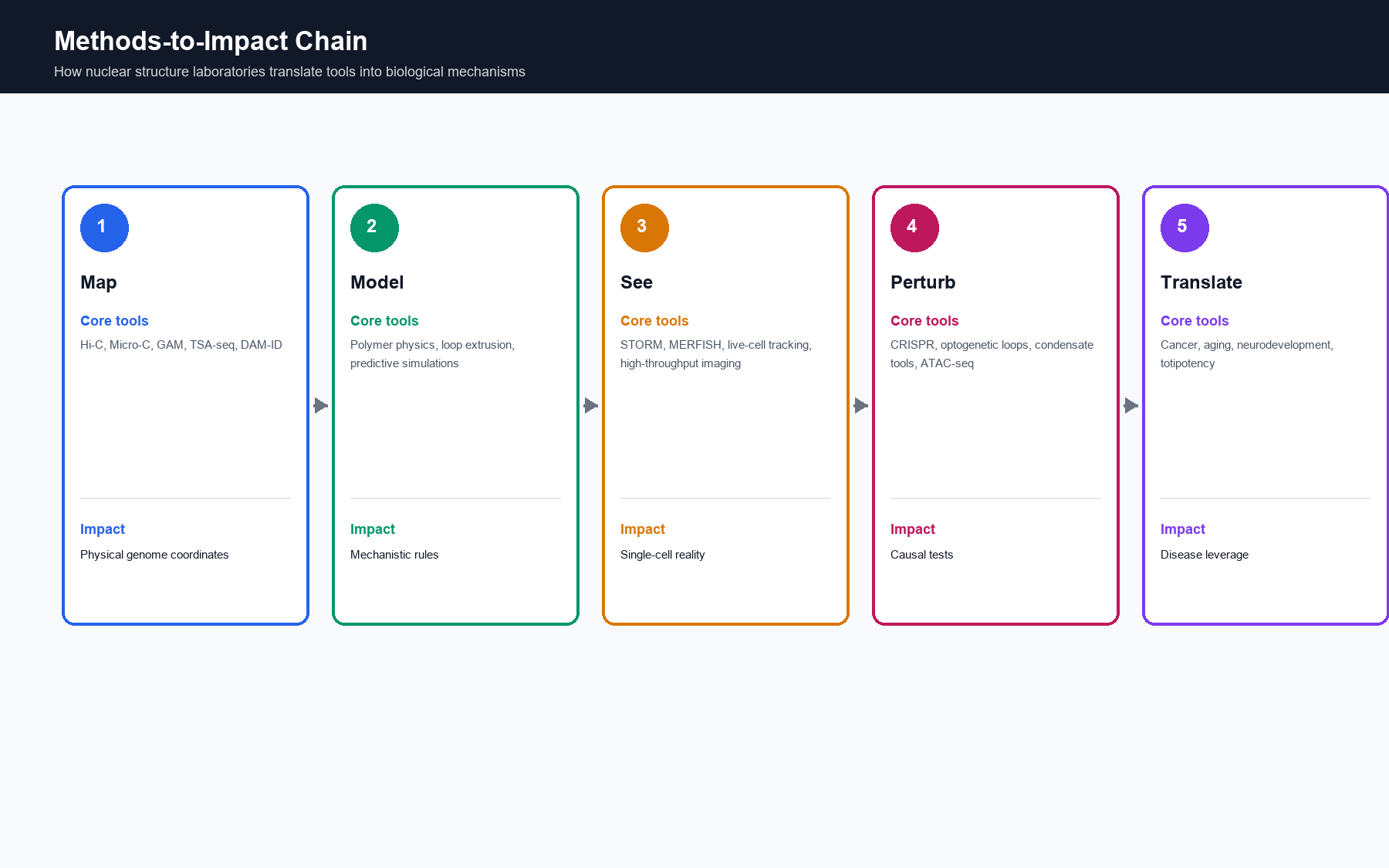
The shared pattern across the document is not simply discovery of more nuclear structures. It is a methods-to-impact chain: quantitative maps made spatial hypotheses testable, physics converted correlations into rules, imaging exposed single-cell reality, perturbation tools made causality possible, and disease systems made architecture actionable.

Map

Hi-C, Micro-C, GAM, TSA-seq, DamID, and single-cell atlases created coordinates for the genome inside nuclei.

Model

Loop extrusion, polymer physics, condensate theory, and chromatin network models made nuclear architecture mechanistic.

Infographic showing how nuclear structure methods translate into biological impact

Spatial maps became the baseline

Genome architecture is now measured as a physical state, not inferred only from linear sequence or gene lists.

Compartments gained material rules

Nucleoli, speckles, paraspeckles, heterochromatin, and transcription hubs can be discussed in terms of assembly, dynamics, and function.

Disease became architectural

Cancer, aging, neurodevelopment, and totipotency now have testable spatial-genome mechanisms, not only altered gene-expression signatures.

Laboratory directory

Contributions and official links

Each profile below condenses the document's contribution summary into a site-ready research-impact note. Homepage buttons point to official lab pages or, where the lab is hosted inside an institutional profile, the most stable official group page.

1. Topographers: mapping the 3D genome

Hi-C, compartments, 4D Nucleome

Job Dekker

University of Massachusetts Chan Medical School

Introduced chromosome-conformation methods that made genome folding measurable at whole-genome scale, anchoring A/B compartments, time-resolved nuclear organization, and high-resolution Hi-C/Micro-C workflows.

Dekker Lab

Fractal globule, Juicer/Juicebox, genome engineering

Erez Lieberman Aiden

Baylor College of Medicine / Rice University

Translated proximity data into interpretable models and shared analysis infrastructure, while using 3D genome engineering to test how architectural boundaries and contacts shape gene regulation.

Aiden Lab

Epigenomics, single-cell chromatin architecture

Bing Ren

UC San Diego / Columbia transition noted on lab site

Linked epigenomic state to 3D folding across development and complex tissues, including allele-resolved folding, droplet Hi-C, and single-cell maps of chromatin regulation.

Ren Lab

GAM, cryoFISH, multi-way contacts

Ana Pombo

Max Delbruck Center for Molecular Medicine

Developed Genome Architecture Mapping to capture genome topology without ligation or crosslinking, revealing multi-way enhancer-promoter hubs and brain-region-specific folding patterns.

Pombo Lab

2. Polymer physics and loop-extrusion mechanics

Loop extrusion, polymer simulations

Leonid Mirny

Massachusetts Institute of Technology

Made chromosome folding a quantitative physics problem, clarifying how loop extrusion, cohesin/condensin dynamics, and compartmentalization compete to generate nuclear architecture.

Mirny Lab

Neurogenome architecture, optogenetic looping

Jennifer Phillips-Cremins

University of Pennsylvania

Connected kilobase-scale chromatin folding to brain function and disease, including repeat-expansion architecture, enhancer logic, and light-controlled perturbation of endogenous loops.

Cremins Laboratory

3. High-resolution imaging and single-cell tracing

STORM, MERFISH, chromatin tracing

Xiaowei Zhuang

Harvard University

Brought molecular-resolution imaging into nuclear structure biology, directly tracing chromosomes and relating epigenetic state to single-cell folding patterns.

Zhuang Research Lab

Live transcription clusters, single-molecule tracking

Ibrahim Cisse

Max Planck Institute of Immunobiology and Epigenetics

Used live-cell single-molecule microscopy to reveal transient Pol II and Mediator clusters, reframing transcription factories as dynamic condensate-like assemblies.

Cisse Laboratory

4. Biomolecular condensates and phase separation

Condensate material properties

Clifford Brangwynne

Princeton University

Helped establish biomolecular condensates as a core organizing principle, with optogenetic tools and nucleolar studies that made phase separation experimentally tractable.

Soft Living Matter Lab

Super-enhancers, transcriptional condensates

Richard Young

Whitehead Institute / MIT

Linked super-enhancer biology to phase-separated transcriptional condensates, RNA Pol II state switching, and selective drug partitioning in cancer-relevant compartments.

Young Lab

Heterochromatin, HP1, remodelers

Geeta Narlikar

University of California, San Francisco

Defines the biochemical rules of heterochromatin and chromatin remodeling, including HP1 phase behavior and rigorous controls for interpreting condensate assays.

Narlikar Lab

5. Nuclear periphery, speckles, and nuclear bodies

Lamina-associated domains, DamID

Bas van Steensel

Netherlands Cancer Institute

Mapped lamina-associated domains and showed how nuclear periphery positioning contributes to repression, reporter behavior, and genome-context effects.

van Steensel Group

TSA-seq, nuclear speckle proximity

Andrew Belmont

University of Illinois Urbana-Champaign

Turned nuclear speckles into measurable genomic landmarks, showing how movement toward speckles can amplify active transcription programs.

Belmont Lab

Architectural lncRNAs, speckles, paraspeckles

David Spector

Cold Spring Harbor Laboratory

Established lncRNAs as structural and disease-relevant organizers of nuclear compartments, including MALAT1, paraspeckles, and breast-cancer organoid models.

Spector Laboratory Profile

Chromosome territories, nuclear positioning

Wendy Bickmore

MRC Human Genetics Unit, University of Edinburgh

Showed that chromosome position and gene relocation are developmentally regulated, with Polycomb compaction and enhancer reach linking nuclear space to cell fate.

Bickmore Group

6. RNA, epigenetics, chromatin dynamics, and reprogramming

lncRNAs, ATAC-seq, ecDNA

Howard Chang

Stanford University

Pioneered noncoding-genome mechanisms and open-chromatin profiling, connecting HOTAIR, ATAC-seq, and ecDNA topology to development, cancer, and aging.

Chang Lab

Xist, X inactivation, Barr body architecture

Edith Heard

EMBL / Francis Crick Institute

Defined chromosome-wide epigenetic silencing as a spatial process, showing how Xist reorganizes X-chromosome topology and how some genes escape inactivation.

Heard Group

Genome cell biology, aging, cancer

Tom Misteli

National Cancer Institute, NIH

Uses imaging and genome cell biology to connect nuclear lamina defects, progeria, cancer, aging, and diagnostic spatial-genome signatures.

Misteli Lab Profile

Histone chaperones, chromatin restoration

Genevieve Almouzni

Institut Curie

Established chromatin as a continuously rebuilt structure, with histone variants and chaperones such as CAF-1 linking DNA replication, repair, and cancer state.

Chromatin Dynamics Team

Totipotency, heterochromatin, transposons

Maria-Elena Torres-Padilla

Helmholtz Munich

Maps how early embryos erase conventional chromatin constraints, using microscopy and single-cell genomics to link nuclear architecture, transposons, and totipotency.

Torres-Padilla Lab

Chromatin networks, fluorescence, modeling

Karsten Rippe

German Cancer Research Center - DKFZ

Integrates single-cell epigenomics, advanced fluorescence microscopy, chromatin editing, and modeling to explain deregulated chromatin networks in cancer.

Chromatin Networks
Editorial scope note

This directory organizes 21 nuclear biology research groups across six thematic domains, summarizing representative experimental and conceptual contributions to nuclear architecture, chromatin folding, nuclear bodies, envelope mechanics, and 3D genome mapping. Official laboratory and institutional links were verified against current institutional directories.

Open ultrastructure atlas