FIB-SEM intake prototype

Nuclear Ultrastructure Atlas

A guided visual page using OpenOrganelle-derived FIB-SEM example figures to explain nuclear envelope context, chromatin texture, nucleoli, nuclear bodies, and cell-type variation.

408
JPEG figures in the intake folder
61
filename-derived visual groups
23
curated page assets
2
MP4 3D reconstructions integrated

Intake analysis

What the figure batch can support

The collection is strongest as an interpretive teaching set. It repeatedly shows segmented or panel-based FIB-SEM views of nuclei from HeLa, Jurkat, macrophage, COS-7, kidney, liver, and immune-contact examples. These exports use darkfield-style contrast: bright structures sit on black backgrounds. The page below uses visible patterns and filename context, but formal figure legends should be checked against the original OpenOrganelle dataset metadata before publication.

Guided readouts

Different aspects of nuclear ultrastructure

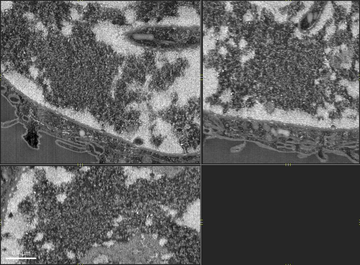
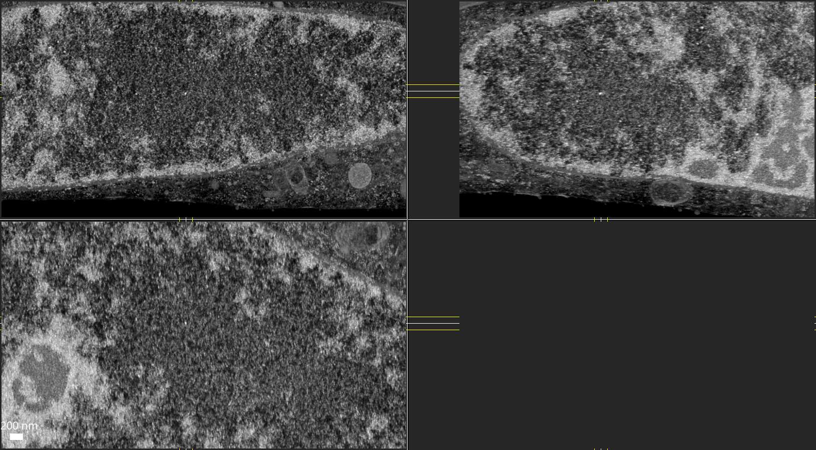
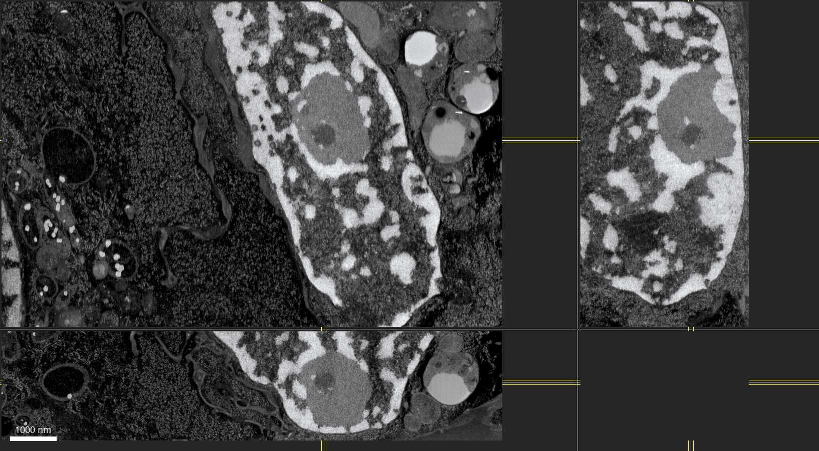
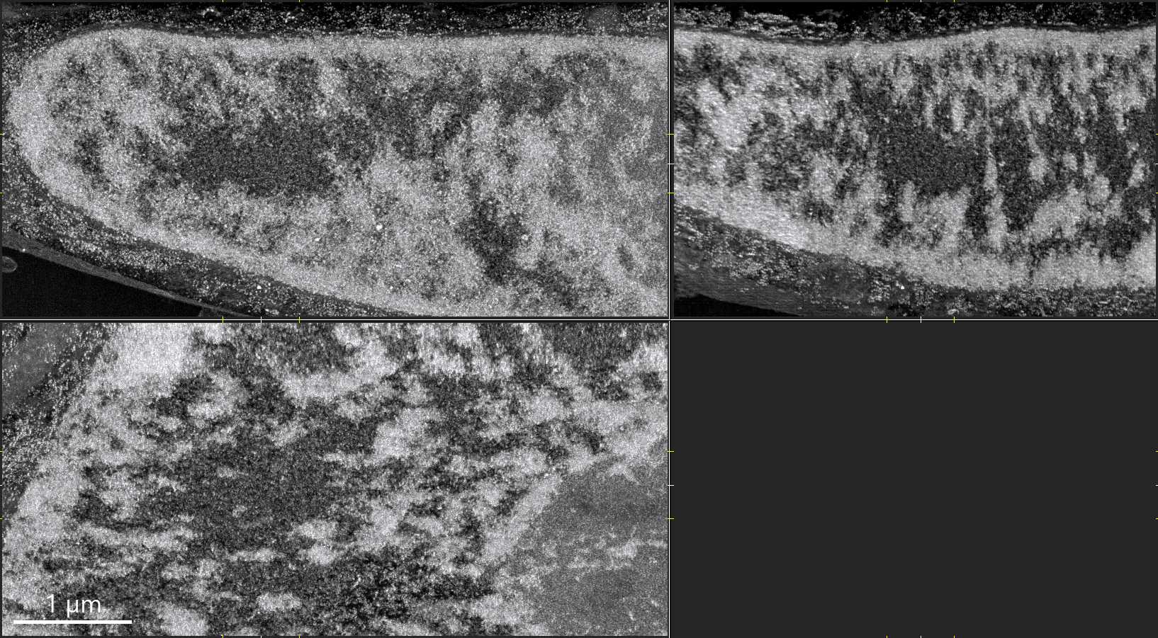
FIB-SEM panels of an interphase HeLa nucleus

Envelope and periphery

The nucleus as a compartment, not a flat outline

FIB-SEM sections place the nuclear boundary inside the surrounding cytoplasm, making the envelope, adjacent organelles, and peripheral chromatin context easier to discuss together. This is useful for explaining why nuclear architecture is both a genome-organization problem and a cell-geometry problem. Volume EM atlas study.

Related nuclear envelope review
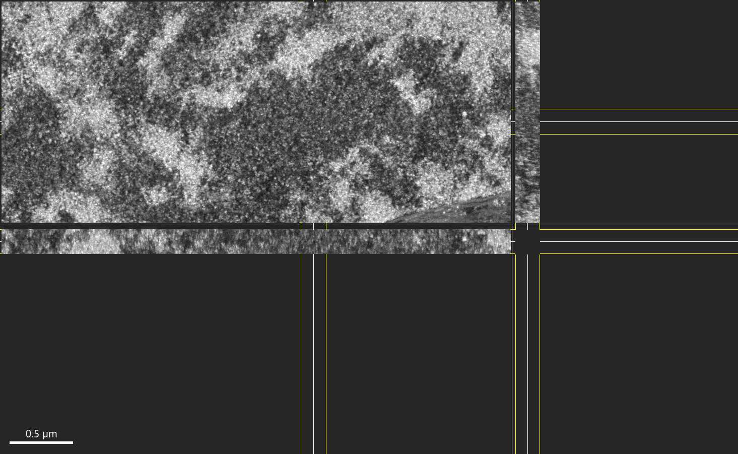
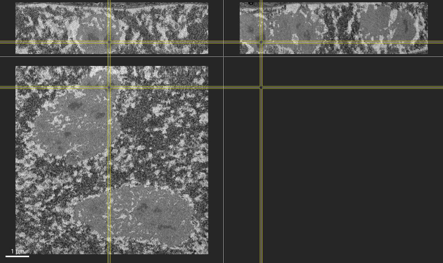
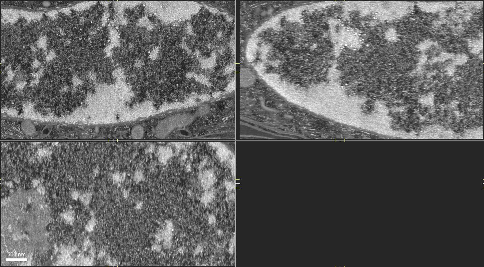
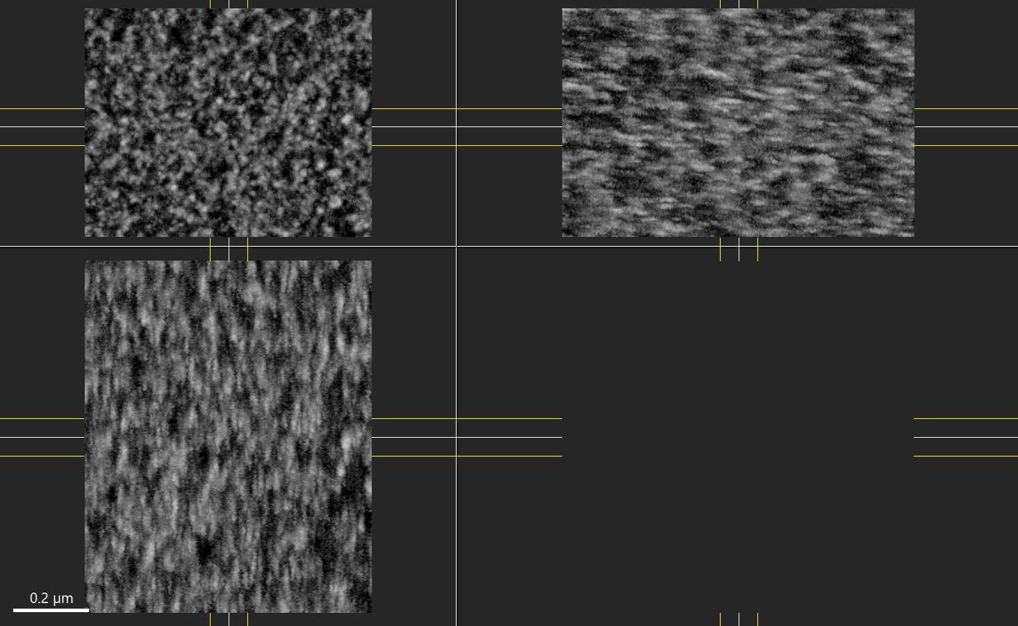
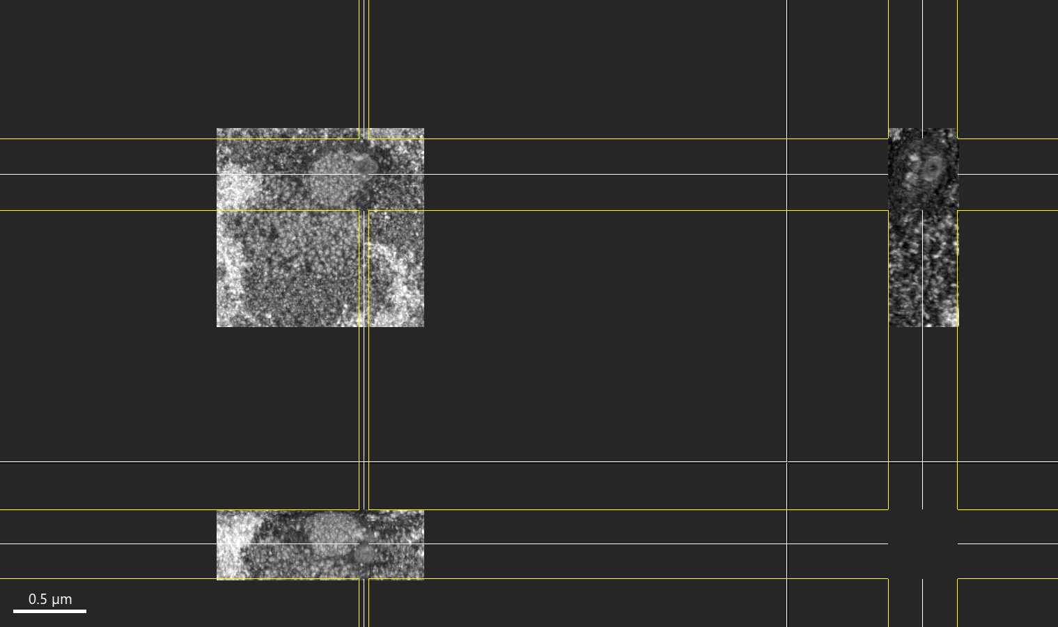
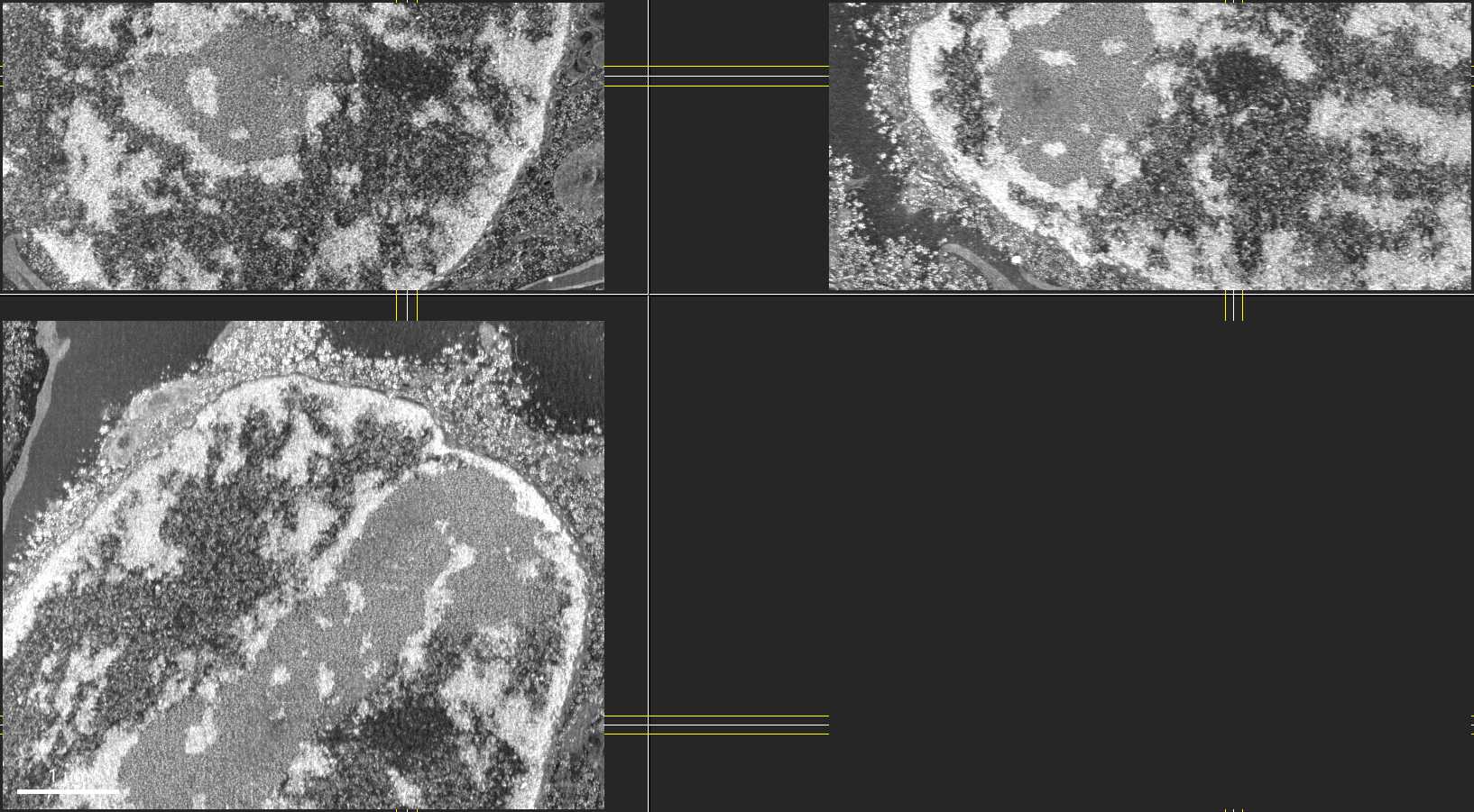
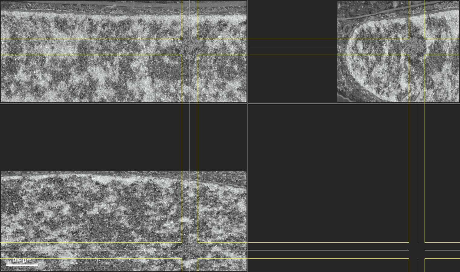
High-magnification FIB-SEM panels showing chromatin texture in an interphase HeLa nucleus

Chromatin texture

Dense and open nuclear material at nanometer scale

The high-magnification panels show heterogeneous electron density inside the nucleus. Used carefully, these views help introduce the visual difference between compacted material, less-dense regions, and the problem of mapping EM contrast onto molecular chromatin states. Volume EM atlas study. Primary segmentation study.

Related heterochromatin review
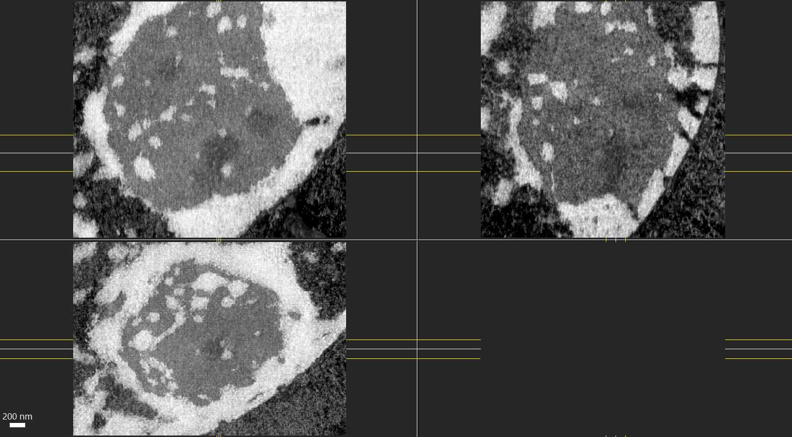
Macrophage nucleus panels showing nuclear body slices
Macrophage nucleus panels showing nuclear body slices without rendered surfaces

Nuclear bodies

Compartment boundaries can be inferred, segmented, and debated

The macrophage examples are useful for showing how the same region can be presented as raw slices or as a more segmented visual. That contrast supports a stronger teaching point: nuclear body interpretation depends on imaging, annotation, and biological context. Primary segmentation study.

Explore nuclear body reviews

Nanoscale Visualization

3D Volume EM Video Explorer

Explore rotation and slice movies of fixed-cell volume EM exports labeled as pancreatic islet and T-cell examples. Rotation and Z-slice traversal are display motions; they do not measure biological dynamics. Dataset identities and annotations still need to be matched to the original records. Volume EM atlas study.

Active: Pancreas Islet Chromatin
Speed:

Select Dataset

Dataset Insight

Pancreas Islet Chromatin (FIB-SEM 3D): This rotation movie shows a rendered peripheral dense region and less-dense channels in a nuclear volume. EM morphology alone does not establish heterochromatin identity, transcriptional activity, or correspondence to nuclear pores; those interpretations require source annotations or correlative markers.

  • Rendered contrast depends on image processing and segmentation.
  • Apparent continuity must be checked against source slices and segmentation.
  • Illustrative morphology; quantitative model calibration has not been verified.

Gemini Flash triage

Model-assisted structure candidates

Model-generated labels and filenames are hypotheses for expert review, not validated annotations. None of the molecular identities below is confirmed by this page. Match each crop to its original dataset and annotation, and use correlative markers where identity is not resolved by ultrastructure. Primary segmentation study.

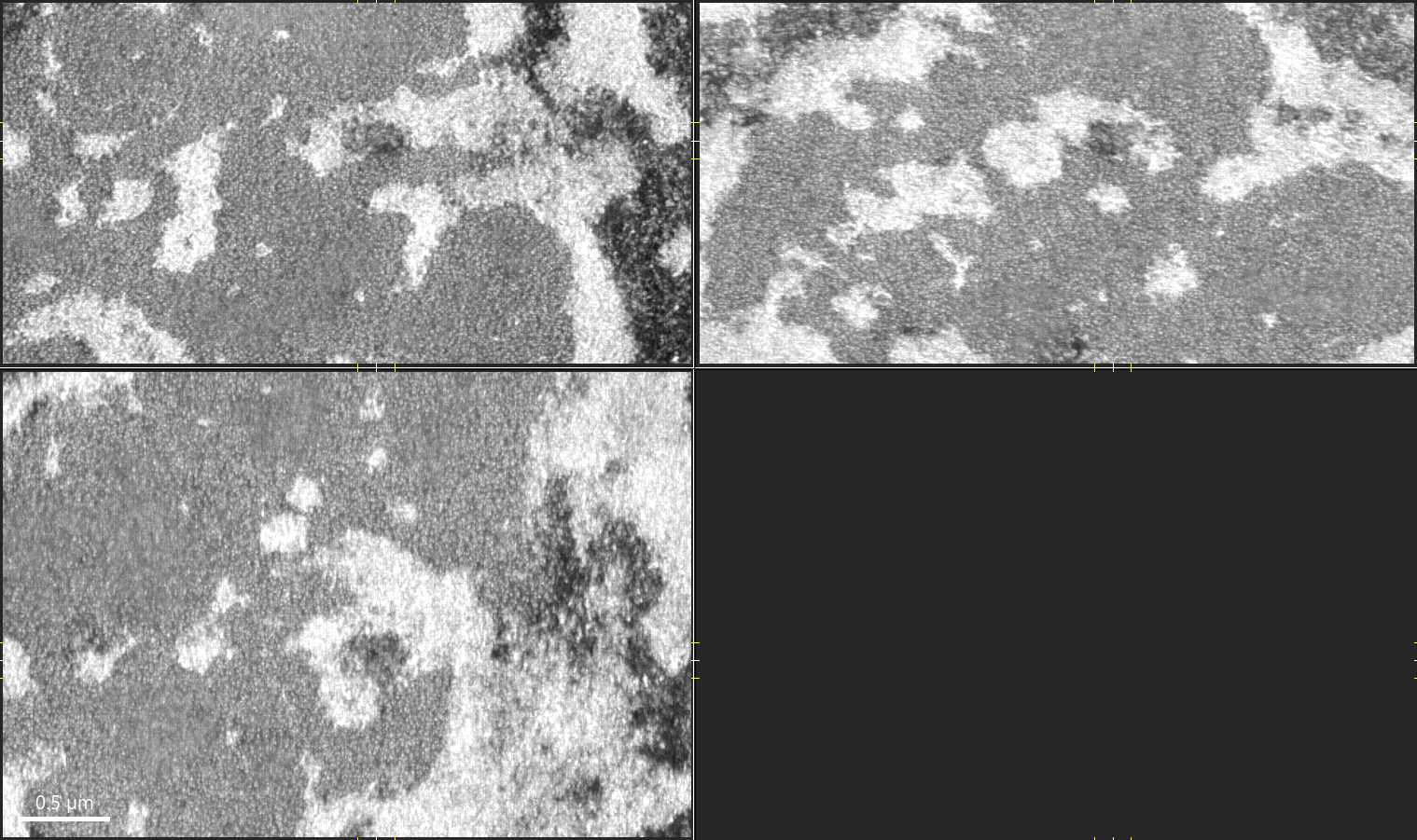
HeLa interchromatin granule cluster candidate from Gemini Flash triage

Unverified candidate

IGC / speckle candidate

The crop shows granule-like contrast. A filename or model label does not confirm an IGC or nuclear-speckle identity.

HeLa candidate Cajal body and nuclear speckle region from Gemini Flash triage

Unverified candidate

Candidate Cajal body

Gemini flagged a discrete textured body near an IGC-like region, but the call depends on the SUBZCB label.

Nucleolus subcompartment candidate from Gemini Flash triage

Unverified candidate

Nucleolus substructure candidate

A nucleolus-like crop with internal contrast variation; fibrillar-center, dense-fibrillar and granular-component assignments require source or expert confirmation.

Macrophage nuclear envelope and heterochromatin candidate from Gemini Flash triage

Unverified candidate

Envelope / dense chromatin candidates

A clear nuclear boundary with coarse dense material at the periphery and throughout the nucleoplasm.

Dense nuclear subregion in a macrophage-labeled export; Polycomb identity unverified

Unverified candidate

Dense-region candidate

A dense subregion in an export whose filename includes PcG. Polycomb proteins, histone modifications and silencing activity cannot be identified from this morphology alone; the original annotation and molecular evidence remain unverified.

Candidate map from Gemini Flash

Candidates requiring verification

  • IGC / nuclear speckles: hela-igc-gamma.jpg, hela-igc-interphase.jpg, grivet-igc-sars-context.jpg, mouse-liver-igc-render.jpg.
  • Nucleolus with DFC/FC/GC candidates: tcell-nucleolus-subcompartments.jpg, hela-nucleolus-subcompartments.jpg, jurkat-nucleolus-subcompartments.jpg.
  • Nuclear envelope / heterochromatin: macrophage and interphase HeLa views were the strongest page-ready examples.

Use with caution

  • Cajal body: Gemini returned medium-confidence calls for hela-cajal-body-candidate.jpg and hela-cajal-body-candidate-render.jpg. These should be labeled as candidates only until checked against the original OpenOrganelle annotation or a human EM review.
  • Nucleolus subcompartments: the DFC, FC, and granular component are most plausible in the nucleolus-labeled crops, but the model cannot confirm molecular identity from contrast alone.

Cell-type variation

Nuclear ultrastructure changes with cellular context

The selected batch includes immune cells, epithelial-like cells, tissue sections, and infection or contact contexts. The safest public framing is comparative: the images show that nuclear shape, surrounding cytoplasm, and internal density patterns vary across preparations. Volume EM atlas study.

COS-7 FIB-SEM panels showing nuclear periphery context

COS-7 nuclear periphery

A compact example for discussing nuclear boundary, adjacent membranes, and local chromatin density.

Jurkat nucleus FIB-SEM panels

Jurkat nuclear morphology

A useful contrast against larger adherent-cell examples, especially for immune-cell nuclear shape.

FIB-SEM panels of a T cell and cancer-cell contact example

Immune contact context

A scene-level example for placing nuclear ultrastructure inside a larger cell-cell interaction.

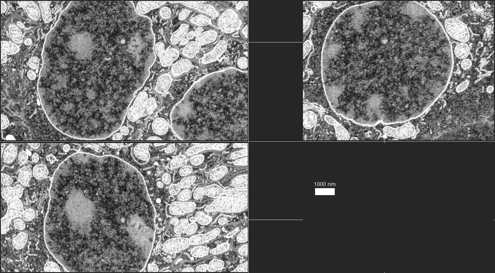
Mouse kidney FIB-SEM panels showing tissue context

Mouse kidney tissue context

A tissue-scale counterpoint for showing how nuclei sit among dense organelle-rich cytoplasm.

Reusable page modules

Assets selected for future pages

Mouse liver FIB-SEM rendering labeled IGC

Interchromatin granule cluster cue

Best used when discussing nuclear speckles or IGC-like domains with clear source confirmation.

mouse-liver-igc-render.jpg
HeLa cargo particle FIB-SEM panels

Particle-rich nuclear regions

A practical supporting image for transport, cargo, or punctate nuclear structure explainers.

hela-cargo-particles.jpg
Macrophage nuclear body slice panels

Nuclear body slice stack

Strongest reusable candidate for explaining raw slices versus annotated nuclear compartments.

macrophage-nuclear-body-slices.jpg
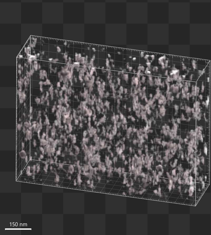
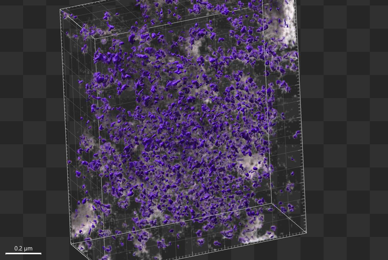
HeLa cell 3 chromatin 3D surface render

3D Surface Chromatin Map

Model-assisted surface rendering of nuclear contrast; genomic domain boundaries and molecular identity have not been verified.

hela-cell3-chromatin-3d.jpg
Pancreas islet chromatin ultrastructure overview

Pancreatic Chromatin Overview

A pancreatic-islet-labeled export showing dense nuclear regions; heterochromatin identity requires source confirmation.

pancreas-islet-chromatin-overview.jpg

Credit and evidence

Source notes

This page is an intake prototype using local example images collected from the OpenOrganelle public resource. Individual image-to-dataset provenance, molecular annotations, and reuse licenses remain unresolved. The linked atlas and segmentation studies describe methods; they do not authenticate every local crop or model-generated label.

OpenOrganelle portal

OpenOrganelle provides public visualization access to cloud-hosted FIB-SEM datasets through the CellMap data portal.

openorganelle.org

Volume EM atlas

Xu and colleagues described an open-access FIB-SEM atlas of whole cells and tissues, with datasets deposited to OpenOrganelle. Volume EM atlas study.

Nature 2021 atlas DOI

Organelle segmentation

Heinrich and colleagues reported segmentation of many organelle classes in 4 nm and 8 nm voxel FIB-SEM volumes. Primary segmentation study.

Nature 2021 segmentation DOI

Portal implementation

The public OpenOrganelle code repository describes the app as a gateway for visualizing large cloud-hosted FIB-SEM datasets.

Janelia CellMap GitHub

Publication record update: 12 October 2026

Scite publication metadata independently confirms Xu et al., the volume EM atlas and Heinrich et al., whole-cell organelle segmentation as separate Nature papers. These method papers establish the resource and reported segmentation work; they do not validate every local crop, model-generated label or molecular assignment.

The atlas has a publisher correction, whose notice was read directly. It replaces a damaged Supplementary Video 2 and corrects axis-label formatting in Extended Data Fig. 2. It does not supply missing provenance for the local example movies. Use the corrected publisher supplements when comparing the original data.

This update checks publication identities and the correction notice. Original full Methods, all dataset annotations, raw-volume segmentation accuracy and individual image reuse permissions remain outside the verified scope. Rotation and slice traversal are display motions of fixed-cell volumes.