CellNucleus.com

Researcher Resources

Microscopy Imaging Software

The commercial software a cell-nucleus researcher actually chooses among for 3D and 4D image analysis — dedicated analysis platforms, microscope-vendor suites, high-content screening software, and a deconvolution specialist — with the open-source tools most laboratories run alongside them.

Entries are drawn from each vendor's own published product pages, consulted in November 2026. Where a vendor's pages do not state a capability or a licence term, this page says "not documented" rather than treating the capability as absent. One entry, GnoCyte, is the site publisher's own software and is labelled as such where it appears.

GnoCyte by Gnometrix Labs

Explore GnoCyte

Science software by Gnometrix Labs

GnoCyte

From microscopy images to quantitative insight: explore multichannel volumes and time series, inspect segmented objects in 3D, and connect spatial measurements with the underlying image.

GnoCyte brings visualization, segmentation review, tracking and quantitative biophysics into a research workspace designed for cell and nuclear imaging.

Explore GnoCyte
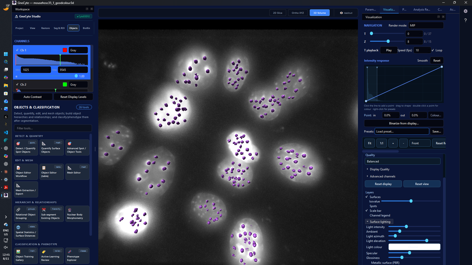
GnoCyte 3D volume workspace showing purple object surfaces over a grayscale microscopy volume.
GnoCyte 3D volume view with object surfaces over microscopy data. Display example; object identity and segmentation accuracy require independent review.

Disclosure: GnoCyte is developed by Gnometrix Labs, which also owns and operates CellNucleus.com. GnoCyte is in development and is not yet on sale.

Explore 3D and 4D data

Inspect multichannel microscopy as slices or 3D volumes and navigate time series. Development work includes out-of-core streaming for large datasets and support for formats such as OME-TIFF, Imaris IMS and OME-Zarr/NGFF.

Segment and measure

Create and review candidate masks, visualize object surfaces, and examine object measurements in the context of the source image. Classical and machine-learning workflows are part of the development scope; model availability and setup vary by build.

Follow nuclear dynamics

Guided GnoCyte and companion GnoCondensate workflows support nuclear and condensate time-course review, including track inspection, nuclear alignment and review of candidate fusion or fission events.

Connect analysis and context

Explore specialist analyses such as FRAP and particle tracking, linked plots, and exportable results. Physical calibration, sampling and model assumptions determine which measurements are interpretable. Local agent integration is also in development.

GnoCyte is research software in development. Confirm the features, file readers, models and GPU support in the particular build being evaluated.

Curated list

Dedicated 3D/4D Analysis Platforms

6 resources

Analysis-first packages, independent of any one microscope brand. These are the established reference points for volumetric rendering, object detection, tracking, and measurement of nuclear and cellular structures in 3D and over time.

GnoCyte — Gnometrix Labs

In development · Publisher's own software

A microscopy research workspace for examining multichannel 3D/4D images, reviewing segmentation and object measurements, and exploring nuclear and condensate dynamics.

  • Slice and GPU volume views, with development support for streaming large datasets and reading open microscopy formats
  • Candidate object segmentation, 3D surface visualization, measurements and linked analysis
  • Guided nuclear and condensate time-course review, with tracking, alignment and candidate event inspection through GnoCyte and companion GnoCondensate workflows
  • Specialist biophysics workflows and research exports; interpretation depends on calibration, sampling and the analysis model

Windows development builds; features, optional models and GPU requirements vary. Not yet on sale.

Explore GnoCyte

Imaris — Oxford Instruments

Version 11.1 · Permanent licence with renewable maintenance plan

Interactive 3D/4D microscopy visualisation and analysis, sold in modular application packages for cell biology, core facilities, and neuroscience.

  • Volume rendering and analysis of very large datasets in its multi-resolution HDF5-based .ims format, with navigation of gigabyte-to-terabyte data
  • Four detection models — Spots, Surfaces, Cells, and Filaments — covering nuclei, cells, neurons, and vessels, plus tracking and colocalisation
  • Batchable, step-by-step Workflows intended to make analyses reproducible across datasets
  • Integrated stitching and ClearView-GPU deconvolution within the same package
  • Scripting through ImarisXT extensions in Python and MATLAB; Windows and macOS
Visit vendor site

ZEISS arivis Pro — ZEISS

Version 4.6 · Subscription licences documented; maintenance agreement also offered

Pipeline-based analysis and GPU-powered 3D/4D visualisation of large, multi-channel, multidimensional datasets; formerly arivis Vision4D.

  • No-code analysis pipelines combining conventional operators and AI models, reusable across datasets and scalable to arivis Hub
  • Converts more than 30 input formats to its Scalable Image Storage (SIS) format for work with large files
  • Deep-learning instance segmentation, including Cellpose and StarDist models and, in 4.6, externally trained 3D ONNX models
  • Tracking and lineage analysis, neuron tracing, and well-plate (high-content) analysis
  • Python operators and scripting; optional VR toolkit for proofreading results; Windows only
Visit vendor site

Amira — Thermo Fisher Scientific

Version 2025.1 · Yearly subscription programme documented for academic and government users

Visualisation and analysis for 2D–5D life-science and biomedical imaging across optical and electron microscopy, CT, and MRI; the sister product Avizo addresses materials science.

  • Multi-modal, multi-scale data handling with Bio-Formats compatibility and support for very large datasets
  • AI-driven segmentation workroom and AI denoising, with more than 200 built-in measurements
  • Image Recipes record workflows — including AI-based ones — for replay without programming
  • Meshing and modelling tools that turn labelled 3D images into inputs for simulation
  • Python and Tcl scripting, a MATLAB bridge, and custom C++ modules; Windows and Linux
Visit vendor site

Aivia — Leica Microsystems

Version 16 · Subscription

An analysis-only platform with an AI-first design, for 2D-to-5D image visualisation, analysis, and interpretation; it does not drive a microscope and accepts images from any imaging system.

  • Ships with 22 applications and 20 pre-trained deep-learning models for segmentation, restoration, and virtual staining
  • Segment by Example (version 16) segments cells in 2D and 3D from as few as three examples
  • Deep-learning models can be trained and applied locally or through AiviaCloud; the Apex plan accepts user-supplied models
  • Reads more than 45 microscopy file formats; desktop application plus browser access through AiviaWeb
  • Python plugins in all plans; Windows desktop
Visit vendor site

Volocity — Quorum Technologies

Version 7 · Licence terms not documented on the vendor's pages

A long-established 3D/4D suite spanning acquisition, volume visualisation, restoration, and quantitation of fluorescence images. Quorum Technologies acquired Volocity from PerkinElmer in 2018; it is no longer a PerkinElmer/Revvity product.

  • Integrated product family: restoration, visualisation, and quantitation modules usable independently or combined
  • Interactive high-resolution volume rendering of multi-channel 3D/4D data
  • Quantitation covering morphology, colocalisation, FRAP, FRET, and ratiometric analysis
  • Hardware-agnostic import, including OME-TIFF, from widefield, confocal, and high-content systems
  • Volocity 7 runs on Windows 10/11; macOS is supported only by version 6.3 and earlier
Visit vendor site

Curated list

Microscope-Vendor Platforms

3 resources

The software supplied with the major microscope brands combines instrument control and acquisition with processing and analysis. For many laboratories this is the default environment; its analysis depth varies by package and module.

NIS-Elements — Nikon

Current 6.x series · Licence terms not documented; NIS-Elements LE is free

Nikon's universal platform for image acquisition, analysis, visualisation, and data sharing across its widefield, confocal, and super-resolution systems.

  • Packages tiered by use, from the free LE edition through BR and the confocal C package to the flagship AR and high-content HC packages
  • NIS.ai modules use convolutional neural networks for segmentation (Segment.ai), denoising, and clarity enhancement (Clarify.ai)
  • 2D and 3D deconvolution available as an option in the AR, C, and HC packages
  • Macro creation in all paid packages, and an NIS.API for integrating external schedulers, robotics, and liquid handlers over HTTP
  • Windows only
Visit vendor site

ZEN — ZEISS

Version 3.14 · Perpetual and subscription (institutional) licences both documented

The universal user interface across ZEISS imaging systems, covering acquisition, processing, 3D visualisation, analysis, and correlative light/electron microscopy; ZEN core extends the same interface to materials and electron microscopy, and ZEN lite is a free edition.

  • More than 180 image-processing tools, batch processing, and a GPU-powered 3D engine presented with very large datasets
  • Deconvolution Toolkit with 15 published algorithms; AI Toolkit with trainable deep-learning segmentation (Intellesis)
  • Modular Toolkits — acquisition, 2D, 3D, AI, Connect, Developer, Bio Applications — licensed per installation
  • The CZI format is fully documented and Bio-Formats compatible, with open-source libCZI libraries; export to OME-TIFF
  • Developer Toolkit provides a Python script editor for acquisition, processing, and analysis; Windows only
Visit vendor site

LAS X — Leica Microsystems

Release tracks per system family · Licence terms not documented; modular licences on LAS X Core

Leica Application Suite X is a single platform to set up experiments, acquire images from Leica microscopes and cameras, and analyse the results; LAS X Office is an acquisition-free variant for review and analysis.

  • LAS X Navigator builds a whole-sample overview for region-of-interest definition and automated high-resolution acquisition
  • Guided, wizard-based analysis workflows, including simultaneous multi-aspect measurements such as spots per nucleus
  • Fully integrated deconvolution using an adaptive calculated or supplied PSF (optional module)
  • Optional modules add time-lapse, Z-series, stitching, high-content screening, FRAP/FRET, colocalisation, and 3D visualisation
  • Deep-learning segmentation is not documented for LAS X itself; Leica documents it in the separate Aivia platform above. Windows only
Visit vendor site

Curated list

High-Content Screening

1 resource

MetaXpress — Molecular Devices

Version 6.7 (classic) · Licence terms not documented on the vendor's pages

High-content image acquisition and analysis software for the ImageXpress instrument family, built around orchestrated plate-based workflows.

  • Turnkey Application Modules cover hundreds of routine assays, and a Custom Module Editor builds multi-step analyses
  • A 3D Image Analysis Module measures volume, intensity, and distances in spheroids, microtissues, and cells in 3D matrices
  • Optional PowerCore parallel processing and the included MDCStore data manager handle plate-scale data
  • A 2D real-time deconvolution option uses the AutoQuant algorithm
  • Deep learning sits in the companion IN Carta software (SINAP segmentation), which the newer HCS.ai systems pair with MetaXpress Acquire. The classic MetaMorph package was discontinued in 2023. Windows only
Visit vendor site

Curated list

Deconvolution Specialist

1 resource

Huygens — Scientific Volume Imaging (SVI)

Version 26.04 · Licence ownership or monthly subscription

A suite built around fluorescence image deconvolution and restoration, with interactive analysis and volume visualisation in 2D–4D; editions include Essential, Professional, Core, and Localizer.

  • Maximum-likelihood deconvolution with theoretical or measured PSFs, by modality: widefield, confocal, spinning disk, STED, light-sheet, and array-detector data
  • Deconvolution Express for automatic restoration and a Workflow Processor for batch deconvolution, stitching, and analysis; GPU and multi-GPU options
  • Object Analyzer adds classic and AI segmentation (including Segment Anything 2 and Cellpose), colocalisation analysis, and object tracking on restored data
  • Reads and writes OME-TIFF and HDF5, including BigDataViewer-compatible export
  • The only package in this guide documented on all three desktop platforms: Windows, macOS, and Linux. Tcl and Python scripting
Visit vendor site

Side by side

Comparison Table

"Not documented" means the vendor's own published pages consulted in November 2026 did not state the capability or term; it is not evidence that the capability is absent. GnoCyte is under development; listed features and platform support may vary by build and system and have not been independently compared here.

Product Primary role Deconvolution Deep-learning / AI segmentation OME-Zarr / NGFF Scripting / API / agent interface Licensing, as the vendor states it Platforms
GnoCyte (in development) 3D/4D analysis with biophysics Not compared Development support; varies by build Development support; varies by build Development interface; varies by build Not yet on sale Windows
Imaris 11.1 3D/4D analysis Yes — ClearView-GPU Machine-learning / AI segmentation documented Not documented (OME-TIFF read) Python/MATLAB XTensions; no agent interface documented Permanent licence with renewable maintenance plan Windows, macOS
ZEISS arivis Pro 4.6 3D/4D analysis pipelines Not documented Yes Not documented (OME-TIFF among 30+ input formats) Python operators and APIs; in-product AI assistant, no external agent interface documented Subscription documented; maintenance agreement also offered Windows
Amira 2025.1 Multi-modal 3D visualisation and analysis Yes Yes Not documented (Bio-Formats compatible) Python, Tcl, C++ modules, MATLAB bridge Yearly subscription programme documented for academic and government users Windows, Linux
Aivia 16 AI image analysis Not documented Yes — central feature Not documented Python plugins; no general API documented Subscription Windows; browser access
Volocity 7 3D/4D acquisition, visualisation, quantitation Iterative Restoration documented Not documented Not documented (OME-TIFF import/export) Not documented Not documented Windows (macOS up to 6.3)
NIS-Elements (6.x) Acquisition and analysis — Nikon systems Yes — option in AR, C, HC packages Yes — NIS.ai CNN modules Not documented Macros; NIS.API over HTTP Not documented (LE edition free) Windows
ZEN 3.14 Acquisition and analysis — ZEISS systems Yes — Deconvolution Toolkit Yes — AI Toolkit Not documented (OME-TIFF export; CZI fully documented) Python (Developer Toolkit) Perpetual and subscription licences documented Windows
LAS X Acquisition and analysis — Leica systems Yes — integrated, optional Not documented for LAS X (see Aivia) Not documented Not documented Not documented (modular licences) Windows
MetaXpress 6.7 High-content screening 2D real-time deconvolution option Not in MetaXpress itself; companion IN Carta provides it Not documented Journals (macros); MDCStore SDK Not documented Windows
Huygens 26.04 Deconvolution and restoration Yes — core function Yes — Object Analyzer (SAM 2, Cellpose) Not documented (OME-TIFF read/write) Tcl and Python; Core compute engine Licence ownership or monthly subscription Windows, macOS, Linux

Context

Open-Source Software Used Alongside

4 resources

Most laboratories pair a commercial platform with open-source tools — for a specialised plugin, a scripted pipeline, or a second opinion on a segmentation. These four are the ones a nucleus researcher is most likely to meet; the Tools page lists further image-analysis resources, including TrackMate, StarDist, and Cellpose.

Fiji / ImageJ

A "batteries-included" distribution of ImageJ with a large community plugin ecosystem, automatic updating, and a macro language; the default tool for everyday image tasks. Licensed under the GNU GPL.

Open resource

napari

A fast, interactive multi-dimensional image viewer for Python (version 0.10.0), built on the scientific Python stack and native Zarr arrays, with layers for labels, points, and tracks and a growing plugin ecosystem. BSD-3-Clause licence.

Open resource

CellProfiler

Pipeline-based quantitative image analysis designed for biologists without programming training (version 4.2.8); saved pipelines make high-content measurements — including nuclear morphology — reproducible at scale. BSD 3-Clause licence.

Open resource

QuPath

Bioimage analysis built for very large whole-slide images (version 0.7.0), with annotation tools, built-in cell and tissue detection, interactive machine learning, and Groovy scripting; widely used for digital pathology research, not clinical diagnosis. GNU GPL v3.

Open resource