PML Bodies
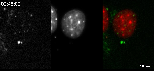
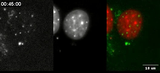
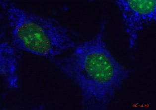
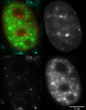
These are timelapse movies of PML bodies (small foci).
The cells are also stained for DNA and/or splicing factor
compartments. Experiments were performed in the Michael J
Hendzel Laboratory in the Department of Oncology at the
University of Alberta.
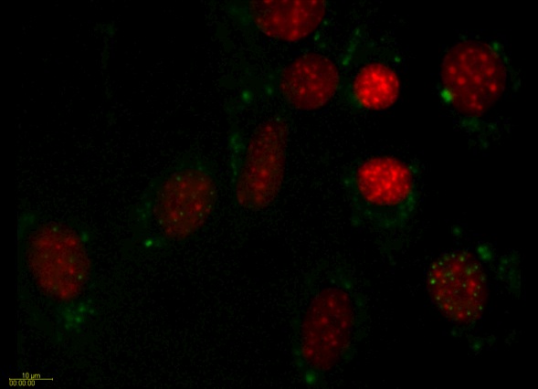
PML Movie 1: SK-N-SH transfected with PML. A field of
SK-N-SH human neuroblastoma cells transfected with
PML-DsRed (green) and counterstained with Hoechst to
visualize the chromatin (red).
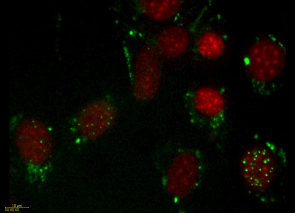
PML Movie 4: SK-N-SH transfected with PML. A field of
SK-N-SH human neuroblastoma cells transfected with
PML-DsRed (green) and counterstained with Hoechst to
visualize the chromatin (red).
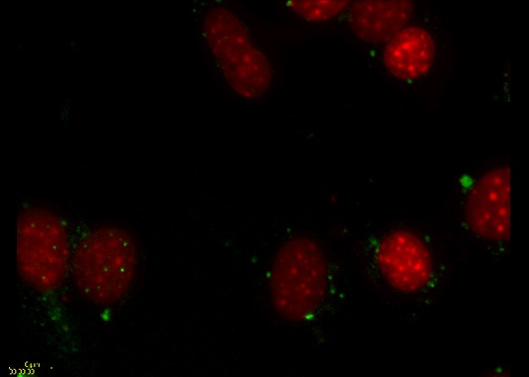
PML Movie 5: SK-N-SH transfected with PML. A field of
SK-N-SH human neuroblastoma cells transfected with
PML-DsRed (green) and counterstained with Hoechst to
visualize the chromatin (red).
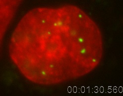
PML Movie 9:SK-N-SH transfected with PML. An SK-N-SH
human neuroblastoma cells transfected with PML-DsRed
(green) and counterstained with Hoechst to visualize
the chromatin (red).

PML Movie 10: SK-N-SH transfected with PML. An SK-N-SH
human neuroblastoma cells transfected with PML-DsRed
(green) and counterstained with Hoechst to visualize
the chromatin (red).
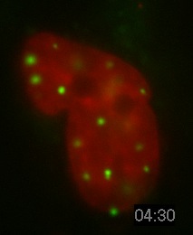
PML Movie 11: SK-N-SH transfected with PML. An SK-N-SH
human neuroblastoma cells transfected with PML-DsRed
(green) and counterstained with Hoechst to visualize
the chromatin (red).
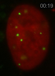
PML Movie 12: SK-N-SH transfected with PML. An SK-N-SH
human neuroblastoma cells transfected with PML-DsRed
(green) and counterstained with Hoechst to visualize
the chromatin (red).

PML Movie 13: MCF-7 transfected with PML. MCF-7 breast
cancer cells transfected with PML-DsRed (red) and
counterstained with Hoechst to visualize the chromatin
(green).
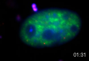
PML Movie 14: A mouse fibroblast cell transfected with
PML Dsred (red), ASF-GFP (splicing factor, green) and
Hoechst (DNA, blue).
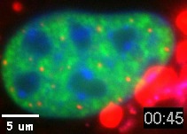
PML Movie 15: A mouse fibroblast cell transfected with
PML Dsred (red), ASF-GFP (splicing factor, green) and
Hoechst (DNA, blue).
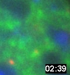
PML Movie 16: A high magnification of a subregion of a
mouse fibroblast cell transfected with PML Dsred (red),
ASF-GFP (splicing factor, green) and Hoechst (DNA,
blue).
