Chromatin Movies
Timelapse images of cell nuclei
illustrating chromatin dynamics. Experiments were performed in the
Michael J Hendzel Laboratory in the Department of Oncology at the
University of Alberta.
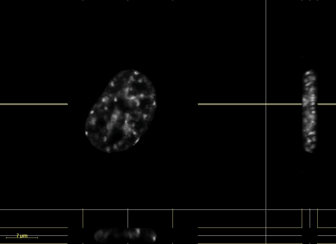
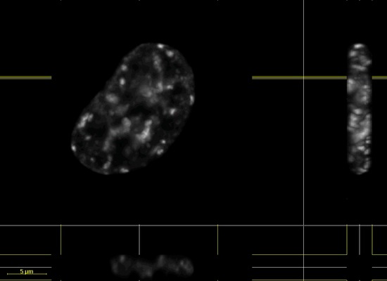
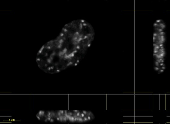
Chromatin Movie 1: An SK-N-SH
cell transfected with DsRed-Histone H1. XY, XZ, and YZ images of a
dataset deconvolved using Huygens (Scientific Volume Imaging)
deconvolution software.
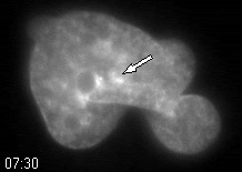
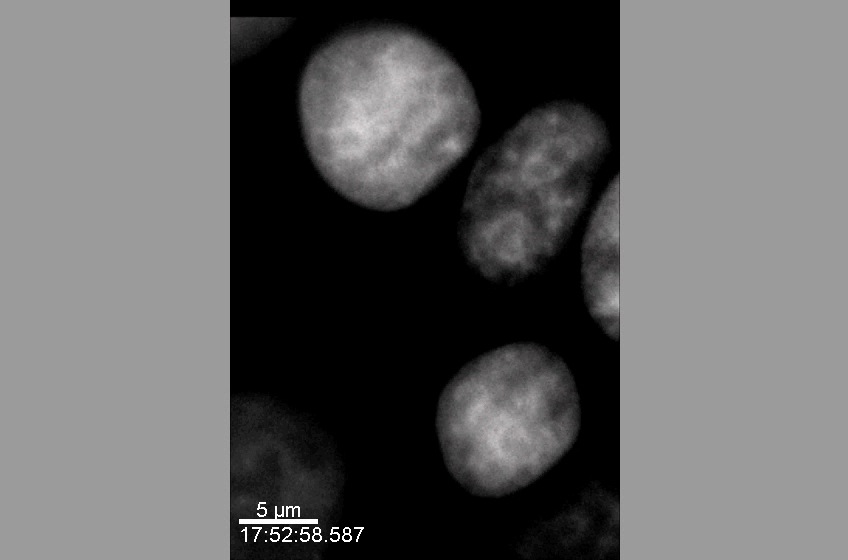
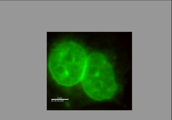
Chromatin Movie 4: A human
glioma cell line (MO59J) stained with Hoechst dye. A 2-D widefield
fluorescence timelapse series is shown.
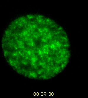
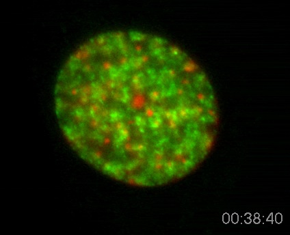
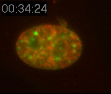
Chromatin Movie 7: A mouse
fibroblast cell showing Rhodamine dCTP-labelled replicated
euchromatin (green) and DNA (red).
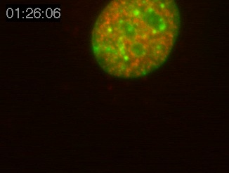
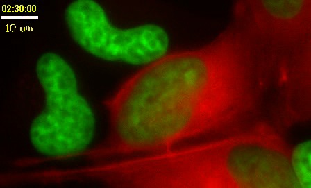
Chromatin Movie 8: A mouse
fibroblast cell showing Rhodamine dCTP-labelled replicated
euchromatin (red) and DNA (green).
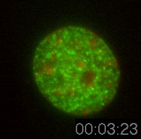
Chromatin Movie 9: A mouse
fibroblast cell showing Rhodamine dCTP-labelled replicated
euchromatin (green) and DNA (red).
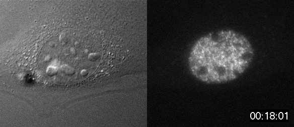
Chromatin Movie 10: A mouse
fibroblast cell showing Rhodamine dCTP labelled euchromatin (right)
and a DIC image (left)
