Diffusion of 40 nm fluorescent
spheres.
Mouse fibroblast nuclei were
microinected with 40 nm fluorescent spheres. In some cases, cells
were also transfected with GFP-SC35 and/or stained for DNA.
Experiments were performed in the Michael J Hendzel Laboratory in
the Department of Oncology at the University of Alberta.
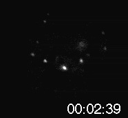
Nanospheres Movie 1.
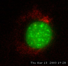
Nanospheres Movie 2: DNA is
green, 40 nm spheres are red.
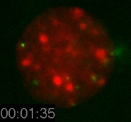
Nanospheres Movie 3: DNA is
red, 40 nm spheres are green.

Nanospheres Movie 4: DNA is
red, 40 nm spheres are green.
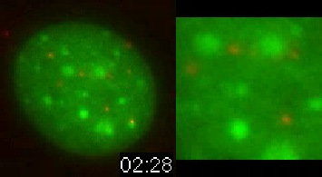
Nanospheres Movie 5: DNA is
green, 40 nm spheres are red.
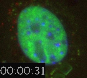
Nanospheres Movie 6: DNA is
blue, 40 nm spheres are red, and SC35-GFP is green.
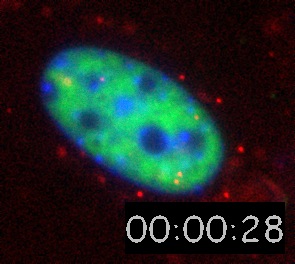
Nanospheres Movie 7: DNA is
blue, 40 nm spheres are red, and SC35-GFP is green.
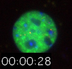
Nanospheres Movie 8: DNA is
blue, 40 nm spheres are red, and SC35-GFP is green.
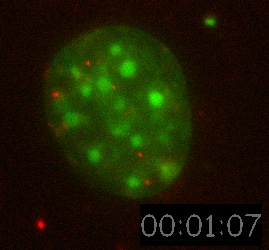
Nanospheres Movie 9: DNA is
green, 40 nm spheres are red.
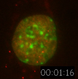
Nanospheres Movie 10: Hoechst
is green, Rhodamine dCTP labelled euchromatin is red. 40 nm spheres
are also red.
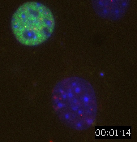
Nanospheres Movie 11: DNA is
blue, 40 nm spheres are red, SC35-GFP is green.
Nanospheres Movie 12: 40 nm
spheres.
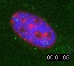
Nanospheres Movie 13: Same as
Nanospheres Movie 8 with the colours changed such that the beads
are green and the DNA is red.
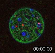
Nanospheres Movie 14: Same as
Nanospheres Movie 8 but filtered using an edge filter to define the
boundaries of structures.
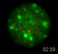
Nanospheres Movie 15: 40 nm
spheres are red, DNA is green.
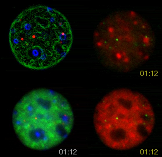
Nanospheres Movie 16: Same as
movie 8 and 14.
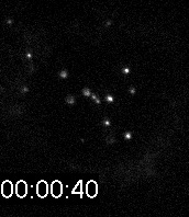
Nanospheres Movie 17: 40 nm
spheres
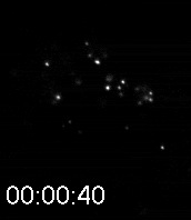
Nanospheres Movie 18: 40 nm
spheres.
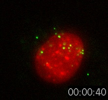
Nanospheres Movie 19: 40 nm
spheres are green, DNA is red.
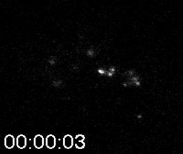
Nanospheres Movie 20: High
speed images. Elapsed time is in min:sec:1/100 sec.
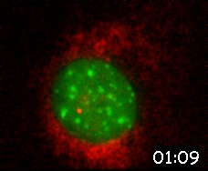
Nanospheres Movie 21: DNA is
green, 40 nm spheres is red. The cytoplasmic signal is
autofluorescence.
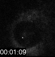
Nanospheres Movie 22: 40 nm
spheres.
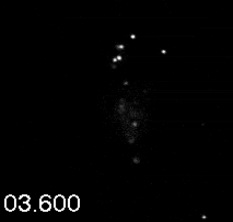
Nanospheres Movie 23: High
speed images. Elapsed time is seconds and 1/10 seconds.


